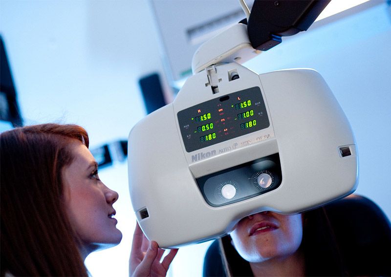
The vision science core facility gives external companies and agencies the opportunities to access the expertise of the vision science staff and equipment to monitor both visual function and ocular structure.
The facility can offer one off assessment and longer term monitoring of both ocular structure and function.
Bioimaging
The group possesses a number of specialised instruments for imaging and measuring the optical properties of the eye. This creates a unique complement of clinical and research tools that include:
Anterior Optical Coherence Tomography (Visante OCT, Zeiss)
Anterior OCT generates cross sectional images of the entire anterior segment of the eye in any plane and allows measurements of the biometrics of the cornea, iris, anterior chamber angle and limbus. This is of interest to researchers who require biometric parameters of the anterior eye, as well as clinicians who require further diagnostic information that may be important for the diagnosis of anterior eye conditions, such as glaucoma, or the need to assess changes after surgery.
Posterior Optical Coherence Tomography (Spectral domain OCT, Heidelberg)
This OCT offers the ability to image cross sections of the eye's posterior segment to monitor change caused by disease or during research trials. Additional software gives the ability to analyse normal and glaucomatous optic discs.
Confocal Scanning Laser Ophthalmoscopy (Heidelberg)
The Heidelberg Retinal Tomograph is a confocal laser scanning system designed for the acquisition and analysis of three dimensional images of the eye's posterior segment. It enables the quantitative assessment of the topography of ocular structures and the precise monitoring of topographic changes.
Corneal Topography (Medmont)
This instrument can measure the outer corneal profile and the results can be linked to those of the Optical Coherence Tomographer to produce a complete optical profile of the anterior eye.
Aberrometry (Imagine-Eyes IRX3)
Measures higher order discrepancies from clear focus that affect all optical systems including the eye. The Imagine-Eyes system uses the most accurate optical components, ensuring precise measurement of the optical aberrations. As the eye is a dynamic system, these higher order aberrations alter over time; these changes can be determined accurately with this method.
Ocular Biometry (IOL Master, Zeiss)
This instrument is designed to accurately measure the axial length of the eye and other biometric data. This information can be linked to corneal topography and anterior OCT measurements to accurately quantify the eye's biometric parameters.
Slit Lamp Biomicroscopes and Anterior Eye Imaging
Used routinely in clinical practice to view and monitor the anterior eye at different angles and to record digital images. The group has a number of slit lamp biomicroscopes with image capture facilities.
Specialised visual function tests
In addition to all standard clinical tests of visual function, the group has specialised instrumentation to measure the characteristics of the ocular media and the retina.
Macular Densitometry (Macular Metrics)
This machine measures the macular pigment optical density at the back of the eye and is the leading instrument in this field as a result of its accuracy, versatility and repeatability in both research and clinical populations. It can be used in studies on nutrition, ageing and pathology.
Light Scattering Meter (CQuant)
The CQuant uses psychophysical techniques to measure intraocular light scatter. It delivers repeatable and simple measures for both clinical practice and research. Changes in light scatter with age or pathology (e.g., cataract) can be assessed and monitored.
The Optometry Clinic
The Optometry Clinic also has access to a broad range of standardised tests (refractive error assessment, central visual acuity, colour vision and peripheral field assessment) and the professional staff available to assess and monitor visual function.
When used in conjunction with bio-imaging both ocular structure and function can be compared over time.

